
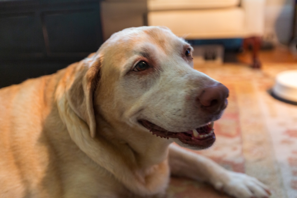
A dog presents with a rectal temperature of 104.8°F, lethargy, and anorexia. The CBC, chemistry panel, and urinalysis reveal nothing definitive. Supportive care resolves the fever within 48 hours, and two weeks later the same dog returns with a 105.2°F temperature and the same nonspecific picture. This is the clinical scenario that defines fever of unknown origin, and it is one of the most diagnostically demanding workups in small animal internal medicine.
For general practitioners navigating FUO cases, the challenge is not recognizing that a systemic process is underway. It is efficiently working through a broad differential in a way that does not exhaust the client’s diagnostic budget before reaching the categories most likely to yield an answer. VESPECON’s clinical advisory services allow general practitioners to engage board-certified internal medicine, oncology, and clinical pathology specialists to work through FUO cases in real time, without the wait associated with formal referral. If your practice is managing recurrent or unresolved fever cases, contact VESPECON about how the partnership model supports these workups.
Key Takeaways
- FUO is defined as a documented core body temperature above 103.5°F on multiple occasions over a period of at least two to three weeks, without an identified cause following a standard initial workup.
- The three major diagnostic categories of FUO in dogs and cats are infectious disease, immune-mediated disease, neoplasia, and a miscellaneous group covering conditions like pancreatitis, panosteitis, and portosystemic shunt.
- Infectious causes are the broadest category and range from focal bacterial infections in deep tissue (pyelonephritis, endocarditis, pyothorax, diskospondylitis) to tick-borne pathogens, systemic mycoses, protozoal disease, and (in cats) FIP, FeLV, and FIV.
- A structured stepwise workup combining signalment, history, physical examination, and targeted laboratory and imaging diagnostics resolves most cases; the threshold for specialist input should be clinical rather than chronologic.
Why Does Fever of Unknown Origin Resist Simple Diagnosis?
FUO resists straightforward diagnosis because the fever itself is a nonspecific endpoint of multiple disease processes that share cytokine signaling but differ entirely in management. The clinician is not trying to confirm the fever; the clinician is trying to identify which diagnostic category is driving it, and those categories produce overlapping presentations at the level of routine bloodwork.
Fever in any species is a cytokine-mediated response to endogenous pyrogens released in the context of inflammation, infection, immune activation, or neoplastic disease. The cytokine signals are produced by macrophages and monocytes responding to a wide range of triggers, and the fever itself provides no information about which trigger is responsible.
The categories also overlap clinically in ways that matter: neoplasia, bacterial infection, and tick-borne disease all can trigger or mimic immune-mediated responses. The stepwise approach to canine fever and the equivalent approach in cats both follow the same underlying logic: signalment and history first, then physical examination, then targeted diagnostics that work systematically through the categories most consistent with the clinical picture.
What Does Signalment and History Tell You First?
Signalment and history narrow the differential dramatically before any laboratory work is run, and a structured patient history is often where the diagnosis announces itself. Age, breed, neuter status, vaccination history, travel and recreational exposure, prior treatment response, and tick history all carry diagnostic weight at the first visit.
Signalment considerations that change the differential:
- Young dogs raise the index of suspicion for panosteitis, hypertrophic osteodystrophy, juvenile cellulitis, steroid-responsive meningitis-arteritis, and congenital portosystemic shunt
- Middle-aged to senior dogs raise the priority of neoplastic causes
- Breed predispositions matter: Cocker Spaniels, English Springer Spaniels, and Miniature Schnauzers for IMHA; Bernese Mountain Dogs, Boxers, and Golden Retrievers for histiocytic and other neoplasias; Beagles and Bernese Mountain Dogs for SRMA; small-breed dogs for MUE
- Outdoor or hunting cats carry elevated risk for FIV, FeLV, bite wound abscesses, and toxoplasmosis exposure
- Cats from multi-cat or shelter environments carry elevated FIP risk
History elements that change the differential:
- Travel history: regional fungal exposure including blastomycosis in the Mississippi and Ohio River valleys, coccidioidomycosis in the desert Southwest, and histoplasmosis along major river drainages
- Recreational exposure: lake swimming or wildlife contact for leptospirosis, brushy or wooded areas for tick exposure, dog parks or boarding for respiratory pathogens
- Prior treatment trials: response or lack of response to antibiotics, anti-inflammatories, or steroids provides meaningful directional information about which category of disease is most likely
- Bite exposure: bite wounds in cats are a leading cause of soft tissue abscess and pyothorax, and the inciting event is often missed on initial history-taking
What Should the Physical Examination Focus On?
The physical examination in an FUO workup needs to be more systematic than a routine examination, because the source of the fever frequently announces itself only when specific structures are evaluated deliberately. Many FUO diagnoses are made on physical examination before advanced diagnostics are needed.
Focus areas drawn from the stepwise approach:
- Lymph node assessment across all peripheral chains, evaluating size, symmetry, texture, and mobility
- Fundic, oral, and otic examinations, which can reveal chorioretinitis (infectious or immune-mediated), oral masses, draining sinus tracts, or otitis media as occult fever sources
- Orthopedic and neurologic assessment, including joint palpation for polyarthritis, paraspinal palpation for diskospondylitis, and gait evaluation for panosteitis or hypertrophic osteodystrophy
- Digital rectal palpation for prostatic disease in intact males and rectal masses
- Spinal and paraspinal palpation for IVDD pain or diskospondylitis
- Careful abdominal palpation for organomegaly, masses, or pain localizing to specific organ systems
- Skin and surface examination for cutaneous lesions, deep tissue swelling, or unhealed wounds that may indicate an abscess or fungal lesion
What Diagnostics Are Needed, and When is Referral Indicated?
The baseline panel for any patient presenting with recurrent or persistent fever of undetermined origin should be considered standard rather than optional. This is what allows the workup to advance to second-tier diagnostics without redundant repeat testing, and stopping short of any component risks missing the early evidence that would have pointed to the correct category.
The components:
- CBC with differential and blood smear review and chemistry panel
- Urinalysis with sediment plus urine culture
- Tick-borne disease panel
- Retroviral testing in cats
More diagnostics are indicated when baseline workup has not produced a diagnosis and the fever pattern persists or progresses, when imaging or sampling of identified lesions becomes the next logical step, and when the differential includes conditions requiring specialist-level management. The threshold for specialist input should be clinical, not chronologic; cases that resist diagnosis are exactly the cases where consultative input shortens the path to answer.
The second-tier workup typically includes:
- Abdominal and thoracic radiographs, including spinal radiographs if paraspinal palpation is abnormal
- Abdominal ultrasound to evaluate splenic architecture, hepatic parenchymal changes, mesenteric and sublumbar lymphadenopathy, and abdominal effusion
- Echocardiography when a new or changing murmur is present
- Urine and blood cultures, including paired cultures when endocarditis is suspected
- Fine needle aspiration of any enlarged lymph nodes, splenic nodules, hepatic lesions, or accessible masses, including aspirates of normal-appearing lymph nodes when hematologic malignancy is on the differential
- PCR screening for infectious organisms when the regional and exposure history support specific pathogens
- Antinuclear antibody testing when systemic lupus is on the differential
- Bone marrow evaluation in patients with unexplained bicytopenia or pancytopenia
When the second-tier workup remains inconclusive, the stepwise approach recommends a directed antibiotic trial (with a different spectrum if a previous trial was unsuccessful) before moving to an empirical corticosteroid trial for suspected immune-mediated disease. The order matters: starting corticosteroids in an undiagnosed infectious case can produce serious deterioration.
For cases where the diagnosis remains elusive after advanced imaging and cytology, or where the clinical picture suggests conditions requiring specialist-level management, VESPECON’s concierge referral service maintains active collaboration with the referring practice throughout the case. VESPECON’s tele-radiology services provide board-certified interpretation of imaging studies with STAT options for cases where abdominal ultrasonography or thoracic radiography findings require specialist-level assessment. Imaging is frequently the step that moves an FUO workup from inconclusive baseline labs to a specific anatomic diagnosis.
What Are the Infectious Causes of FUO?
Infectious disease is the broadest category in the FUO differential and the one most often underestimated when general practitioners triage cases as “probable autoimmune” or “probably cancer” before working through the infectious possibilities systematically. The categories below cover the most common infectious presentations in small animal practice.
Bacterial Infections
Focal bacterial infections in deep tissue produce fever without obvious localizing signs and are among the most clinically rewarding FUO diagnoses, since they are typically treatable once identified.
- Pyelonephritis is one of the most commonly missed infectious causes of fever because the urinary signs that prompt a culture in cystitis are frequently absent. Upper urinary tract infection often presents only with fever, lethargy, and inappetence; a urine culture is appropriate in any FUO workup
- Infectious endocarditis is uncommon but devastating when missed. A new murmur in a febrile patient, or fever in a patient with known valve disease, warrants echocardiography and blood cultures.
- Pyothorax is more common in cats than dogs, often following an unrecognized bite wound or migrating foreign body. Tachypnea, dyspnea, and fever combine in the typical presentation; thoracic imaging and thoracocentesis are diagnostic.
- Soft tissue abscess from bite wounds, foreign body migration, or contaminated trauma can sit deep enough that palpation does not localize it. Cats with apparent FUO and a history of outdoor access often have an unrecognized cat fight abscess as the actual source.
- Diskospondylitis produces fever, spinal pain, and reluctance to move; the diagnosis is radiographic, but the index of suspicion comes from careful spinal palpation.
- Bronchopneumonia may present without a productive cough in early disease; thoracic radiographs belong in any FUO workup where infectious causes are on the differential.
- Prostatitis in intact male dogs can present with fever, lethargy, and caudal abdominal discomfort; rectal palpation and prostatic wash are diagnostic.
Tick-Borne Disease
Tick exposure history is among the most important elements of the FUO workup and among the most frequently underexplored.
- Lyme disease can produce episodic fever with joint pain and lethargy that waxes and wanes for months before a diagnosis is established
- Anaplasmosis targets granulocytes and commonly presents with fever, thrombocytopenia, and lethargy; in some regions, it is the most common cause of acute thrombocytopenia in dogs
- Rocky Mountain spotted fever causes endothelial damage and severe thrombocytopenia with potentially fatal progression if treatment is delayed
- Ehrlichia and Anaplasma species produce overlapping clinical presentations affecting white blood cells and platelets; chronic ehrlichiosis can mimic primary IMT so closely that treatment for the wrong condition is a real clinical risk without testing
Systemic Fungal Disease
Systemic mycoses are regionally distributed but produce FUO consistently enough that travel history matters in every workup.
- Blastomycosis is endemic to the Mississippi and Ohio River valleys and the Great Lakes region; it produces fever, weight loss, respiratory signs, lameness, and ocular disease in dogs
- Coccidioidomycosis (Valley Fever) is endemic to the desert Southwest including Arizona, New Mexico, and parts of California and Texas; respiratory disease, bone pain, and lameness are common presentations alongside fever
- Histoplasmosis is distributed along major river drainages in the central and eastern United States; GI and respiratory presentations predominate
- Cryptococcosis affects cats more frequently than dogs and presents with respiratory or central nervous system disease
Protozoal Disease
- Toxoplasmosis in cats can present with fever alongside ocular disease, neurologic signs, or hepatic involvement; serology and tissue sampling are diagnostic
- Neosporosis in dogs produces neurologic disease, polymyositis, and occasional FUO; serology and PCR are diagnostic
- Babesia species transmitted by ticks cause hemolysis, fever, and thrombocytopenia; co-infection with other tick-borne pathogens is common
Cat-Specific Viral and Inflammatory Disease
- Feline infectious peritonitis is one of the most important FUO differentials in young cats, particularly those from multi-cat or shelter environments; the effusive and non-effusive forms differ in presentation but share persistent fever as a near-universal feature
- Feline leukemia virus can produce fever directly during retroviral infection and indirectly through bone marrow suppression, lymphoma, and opportunistic infections
- Feline immunodeficiency virus predisposes affected cats to opportunistic infections and chronic stomatitis, with persistent low-grade fever a common finding
- Upper respiratory tract infection complexes including feline calicivirus and herpesvirus can produce fever, particularly when chronic shedding or stomatitis is present
- Panleukopenia in unvaccinated cats produces fever, severe leukopenia, and GI signs
Leptospirosis
Leptospirosis deserves separate emphasis given its zoonotic potential and regional resurgence. Dogs with outdoor exposure to standing water or wildlife present with fever, vomiting, polyuria-polydipsia, and azotemia; the spectrum runs from mild self-limiting illness to fulminant hepatic and renal failure.
What Are the Neoplastic Causes of FUO?
Neoplasia should sit near the top of the FUO differential in any middle-aged or older patient with persistent or episodic fever, particularly when the picture includes unexplained weight loss, peripheral lymphadenopathy, or episodic hematologic changes. The presentations that hide longest are also the ones with the most aggressive natural histories, which makes earlier recognition meaningfully different from later recognition.
The hematologic malignancies, particularly lymphoma, lymphoid leukemia, and multiple myeloma, produce fever through sustained immune activation and cytokine release. Feline lymphoma can present with vomiting, weight loss, and fever in a pattern that closely mimics inflammatory bowel disease, and differentiation requires biopsy with histopathology. Canine lymphoma typically presents with peripheral lymphadenopathy, but mediastinal and abdominal forms can present with fever as the primary or only early sign.
Hemangiosarcoma deserves particular attention in the FUO context. Splenic hemangiosarcoma commonly presents with intermittent episodes of weakness, pallor, and fever secondary to intratumoral hemorrhage, with apparent recovery between episodes as the hematoma resorbs. The patient that presents with acute collapse and recovers, then deteriorates episodically over weeks before the diagnosis is established, is a characteristic hemangiosarcoma presentation. Abdominal ultrasound is indicated in any middle-aged or senior dog with episodic fever and anemia.
Osteosarcoma causes localized bone pain, lameness, and sometimes fever in large-breed dogs, and early medullary lesions may not be apparent on initial imaging. The signs of cancer in pets that overlap most with FUO include unexplained weight loss, persistent lethargy, swollen lymph nodes, and appetite changes that persist despite symptomatic treatment.
Other neoplastic causes worth listing on the differential:
- Myeloproliferative disease in cats, frequently FeLV-associated
- Other leukemias beyond lymphoid leukemia, including acute and chronic myeloid leukemias
- Histiocytic sarcoma, particularly in Bernese Mountain Dogs and Flat-Coated Retrievers
- Mast cell tumors, which occasionally release histamine systemically and produce fever and GI signs
- Oral tumors and other tumors with secondary infection or necrosis driving inflammation
What Are the Immune-Mediated Causes of FUO?
Immune-mediated disease drives persistent fever through sustained cytokine release during active hemolysis, ongoing immune complex deposition in tissue, and the inflammatory cascade that accompanies these processes. The distinction between primary immune-mediated disease and infection-triggered secondary immune disease has direct treatment implications, which is why tick-borne disease panels and infectious workup are not optional add-ons in this category.
Immune-mediated hemolytic anemia is one of the most serious immune-mediated conditions in small animal practice, and fever is a consistent component of the active hemolytic phase. The clinical picture of pale or jaundiced gums, tachycardia, tachypnea, and fever in a Cocker Spaniel, English Springer Spaniel, or Miniature Schnauzer is IMHA until ruled out by diagnostics. The breed predisposition in these breeds is well-established, and owners of predisposed individuals should be counseled to treat these signs as prompts for same-day evaluation.
Immune-mediated thrombocytopenia produces fever secondary to the same cytokine cascade, alongside petechiation, ecchymosis, and spontaneous mucosal bleeding when platelet counts drop below critical thresholds. Distinguishing primary IMT from ehrlichiosis-triggered secondary thrombocytopenia requires tick disease testing as a standard component of the workup. Some patients present with concurrent immune-mediated conditions affecting both red blood cells and platelets simultaneously (Evans syndrome), producing fever, pallor, and spontaneous bleeding.
Beyond the hematologic immune disorders, several other immune-mediated conditions deserve specific recognition:
- Immune-mediated polyarthropathy produces fever, shifting limb lameness, and reluctance to move; arthrocentesis with cytology is the diagnostic gold standard, and the condition is frequently missed when palpation does not specifically address each joint
- Systemic lupus erythematosus is uncommon but produces a multisystemic picture with fever, polyarthritis, dermatologic lesions, glomerulopathy, and variable hematologic involvement; ANA testing is the starting point but diagnosis requires meeting defined clinical criteria
- Granulomatous meningoencephalitis and other immune-mediated CNS diseases (collectively MUE) produce fever, neurologic signs, and CSF inflammation; small-breed dogs are overrepresented and MRI plus CSF analysis are diagnostic
- Steroid-responsive meningitis-arteritis (SRMA) in young adult dogs of certain breeds (Beagles, Boxers, Bernese Mountain Dogs) produces fever, neck pain, and reluctance to move; CSF pleocytosis is diagnostic
- Steroid-responsive fever as a category includes cases of persistent fever that respond to corticosteroid therapy without a definitive underlying immune diagnosis ever being established
- Pemphigus foliaceus in cats and dogs produces fever during active immune-mediated dermatologic flares
- Panniculitis and other sterile inflammatory dermatologic conditions can drive systemic fever
- Immune-mediated neutropenia in cats produces fever alongside the increased infection risk that follows neutrophil destruction
What Miscellaneous Causes Belong on the Differential?
A smaller but clinically important group of conditions sits outside the infectious, neoplastic, and immune-mediated categories. These miscellaneous causes are easy to overlook precisely because they don’t fit the major buckets, but they account for a meaningful portion of cases when the workup is not yielding answers in the expected directions.
- Acute pancreatitis produces fever alongside vomiting and abdominal pain in dogs; cats more often present with vague lethargy and inappetence and require specific lipase testing (Spec fPL or cPL) rather than relying on amylase or general chemistry findings
- Portosystemic shunt in young dogs can produce intermittent fever alongside the neurologic and growth abnormalities classically associated with the condition; bile acids and abdominal ultrasound are diagnostic
- Intervertebral disc disease with associated inflammation can produce mild fever alongside spinal pain, particularly during the acute phase of disc extrusion
- Panosteitis in young large-breed dogs produces shifting limb lameness, fever, and reluctance to exercise; the diagnosis is radiographic
- Hypertrophic osteodystrophy in young large-breed dogs produces fever, painful long bone metaphyses, and reluctance to move
- Drug reactions in cats, including penicillin and tetracycline class antibiotics, can produce persistent fever that resolves with discontinuation
Frequently Asked Questions About FUO
What is the most important next step when fever persists beyond two weeks?
A structured second-tier workup including tick disease panel, urine culture, abdominal ultrasound, and cytologic sampling of any identified lesions. Repeating symptomatic management without advancing the diagnostic workup delays the diagnosis and allows underlying disease to progress.
How does the FUO differential differ between dogs and cats?
Cats have a meaningfully different infectious differential, with FIP, FeLV, FIV, upper respiratory complex infections, and toxoplasmosis carrying weight that dog-focused workups can miss. Immune-mediated polyarthritis and SRMA are more common in dogs, while immune-mediated neutropenia and pemphigus foliaceus appear more commonly on the feline differential. Drug reactions, particularly to penicillins and tetracyclines, appear on the feline differential in a way that they do not for dogs.
When does an FUO case warrant referral to a specialist?
When baseline diagnostics and second-tier imaging have not established a diagnosis, when the presentation suggests conditions requiring specialist-level management such as systemic lupus, complex immune-mediated disease, or hematologic malignancy, or when the client needs oncologic staging and treatment planning that exceeds general practice scope. VESPECON’s specialty consultation model provides specialist input at any of these decision points without the full referral process.
Moving FUO From Frustrating to Resolved
Fever of unknown origin is one of the cases where clinical method matters most. The four diagnostic categories that account for most FUO presentations in dogs and cats (infectious disease, immune-mediated disease, neoplasia, and miscellaneous causes) each require a different therapeutic approach, and treating the wrong one has real consequences. A structured workup that moves systematically through these categories, supported by specialist input at appropriate decision points, is what transforms a chronically cycling case into a diagnosis and a plan.
VESPECON’s network of internal medicine, oncology, clinical pathology, and cardiology advisors supports general practitioners through exactly these cases. Reach out to us to learn more about our partnership.
For pet owners: if your pet has had a fever on more than one occasion without an identified cause, ask your veterinarian whether a structured FUO workup through a VESPECON partner practice is the appropriate next step. Most FUO cases have an answer. Finding it requires the right diagnostic approach, and often the right specialist input at the right moment.



Leave A Comment