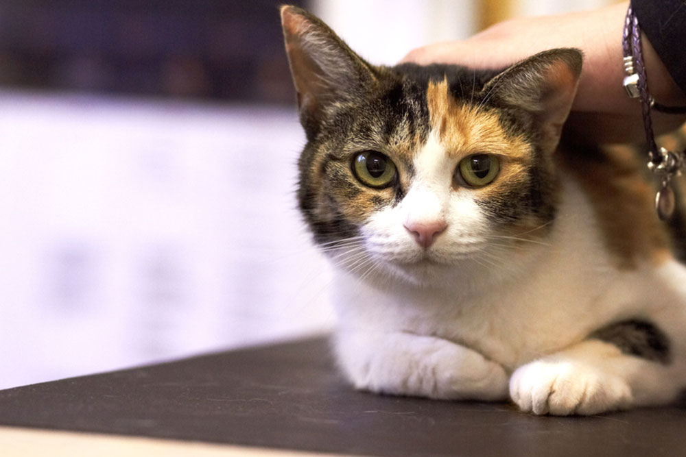
Neurolocalization is one of the highest-leverage skills in small animal medicine, and it is also one of the easiest to short-cut on a busy day. A circling, head-pressing dog and a symmetrically ataxic one can both arrive as “she’s just acting weird,” but the workup, the urgency, and the referral conversation diverge the moment you put a region on the lesion. Get it right and the diagnostics are targeted, the timing fits the condition, and the conversation with the owner is honest about what comes next. Get it wrong and you have either over-spent on a stable case or under-reacted to one where hours matter.
VESPECON’s neurology consultants partner with general practices on exactly this work, providing real-time support that lets you think through a complex case with board-certified input at the point of decision rather than after a workup has stalled. Our specialty consultation services slot into the day-to-day workflow of partner practices, so localization, diagnostic prioritization, and referral timing all benefit from a specialist read at the moment it matters. If your practice sees neurologic cases regularly and you would like steadier scaffolding for handling them, reach out about partnering with us.
Why Neurolocalization Matters in General Practice
- Localization shapes everything downstream: the differential, the diagnostic plan, the urgency call, and the referral decision.
- Mislocalization is expensive: the common errors (vestibular versus cerebellar, upper versus lower motor neuron) drive disproportionate downstream cost.
- One confident approach handles most cases: GPs working from a settled localization keep more cases in-house and refer the right ones at the right time.
- Consultation works best at the decision point: specialist input at localization, not after a workup has stalled, saves time and money.
How Do Neurologic Cases Reach General Practice?
Pet owners rarely describe a neurologic case as such. They describe “wobbly,” “off balance,” “not himself,” “circling,” “weak in the back end,” or “having seizures.” The clinical work begins with translating those owner observations into anatomically meaningful findings and then localizing those findings to a region of the nervous system.
The forebrain, brainstem, cerebellum, vestibular system, spinal cord, and peripheral nervous system each produce characteristic patterns, and misreading the pattern wastes the rest of the workup. A forebrain seizure presentation needs MRI of the brain. A thoracolumbar disc presentation needs imaging of the spinal cord at that segment. Imaging the wrong area produces a normal study that fails to rule out the disease that is actually there.
For pet owners reading along, the discussion that follows is written for veterinary professionals. If you are noticing changes in your pet’s coordination, balance, or behavior, your regular veterinarian is your starting point, and if their practice partners with us, your case may already benefit from specialist input.
Which Localization Patterns Show Up Most in GP?
Most neurologic cases fall into a handful of recognizable patterns, each with its own localization logic.
Seizures and Epilepsy
Seizures are abnormal electrical activity in the brain producing motor, sensory, autonomic, or behavioral signs, classically collapse, paddling, drooling, urination or defecation, and post-ictal confusion. Seizure first response is straightforward: protect the patient from injury, do not restrain, do not place anything in the mouth, and time it. Once a seizure runs past five minutes, or once two or more seizures cluster without recovery in between, you are treating status epilepticus and IV anticonvulsants enter the picture.
The differential depends on signalment and history:
- Idiopathic epilepsy: the diagnosis of exclusion in dogs aged 1 to 6 with normal interictal exams.
- Structural disease: neoplasia, vascular events, or inflammatory disease, more likely outside that age range or alongside abnormal interictal findings.
- Metabolic and toxic causes: hepatic encephalopathy from portosystemic shunts, hypoglycemia, and the toxin-induced seizures in cats.
- Brain neoplasia: a primary consideration in onset over age 6 or with focal signs.
At first presentation, the localization question is forebrain (the likely source given seizure semiology) versus other regions, which are rare for true seizures but worth ruling out on a careful exam.
Cognitive Dysfunction in Aging Pets
Cognitive dysfunction syndrome shows up as disorientation, altered interaction, sleep-wake changes, house-soiling, and anxiety in older patients. The diagnosis is clinical, but the differential deserves real attention, because several treatable conditions look the same from the doorway: hypothyroidism, hypertension, hepatic encephalopathy, brain neoplasia, and chronic pain all mimic it. Localization is generally forebrain, so the work here is less about pinpointing neuroanatomy and more about confirming the diagnosis and ruling out the look-alikes that actually have a fix.
Vestibular Disease and Balance Disorders
Vestibular disease presents with head tilt, circling, nystagmus, and loss of balance, and the critical localization question is peripheral or central. Getting that distinction right is what determines whether the case is managed conservatively or needs emergent imaging.
| Feature | Peripheral vestibular | Central vestibular |
| Nystagmus | Horizontal or rotary, fast phase away from the head tilt | Vertical or direction-changing |
| Mentation | Usually normal | Often altered |
| Proprioception | Intact | Deficits present |
| Common causes | Idiopathic geriatric, otitis media and interna | Stroke, neoplasia, inflammatory, metabolic (Cushing’s, systemic hypertension) |
| Management | Often conservative | Needs emergent imaging |
The classic canine vestibular disease case is the older dog who tilts hard one morning and looks worse than they are, but central disease hides inside that same presentation. Repeat the cranial nerve and proprioception exam if anything feels off.
Degenerative Myelopathy and Progressive Mobility Loss
Degenerative myelopathy is a progressive, painless spinal cord disease primarily of large breeds, presenting as gradual hindlimb ataxia and weakness over months with preserved nociception until late. The challenge is that DM is essentially a diagnosis of exclusion: the same chronic progressive UMN paraparesis can come from intervertebral disc disease, neoplasia, or FCE. SOD1 genetic testing supports the diagnosis in compatible breeds but does not confirm it, and MRI of the thoracolumbar spine remains the way to rule out structural disease.
IVDD and FCE in Sudden Spinal Cord Injuries
Intervertebral disc disease presents as acute back pain, reluctance to move, hunched posture, and varying UMN paraparesis or paraplegia. The grading system tells you the urgency:
- Grade 1 (pain only): conservative management is often appropriate.
- Grade 2 (ambulatory paraparesis): conservative or surgical depending on trajectory.
- Grade 3 (non-ambulatory, voluntary motor): surgical referral indicated.
- Grade 4 (paraplegic, nociception present): urgent surgical referral.
- Grade 5 (paraplegic, absent deep pain): emergent referral, with a recovery window measured in hours.
Fibrocartilaginous embolism presents acutely, often during exercise, and is typically asymmetric, painless, and non-progressive, with most cases recovering substantial function. Once you have a clean localization, the imaging area follows:
| Signs | Localizes to |
| UMN signs to all four limbs | C1-C5 |
| UMN front, LMN rear | C6-T2 |
| UMN rear only, normal forelimbs | T3-L3 |
| LMN signs to rear | L4-S3 |
Wobbler’s Syndrome and Cervical Spinal Cord Compression
Wobbler’s syndrome, or cervical spondylomyelopathy, involves cervical spinal cord compression in large and giant breeds. The presentation is uncoordinated movement, variable neck pain, and generalized weakness that is often more pronounced in the rear than the front despite the cervical localization, which is the part that trips up an exam built on rear-limb signs alone. Management ranges from medical care with activity restriction to surgical decompression, depending on severity and trajectory.

Which Diagnostics Support Neurologic Cases?
Localization narrows the differential before any imaging is ordered, and the localized lesion then drives both the modality and the area:
- Bloodwork: screens for metabolic and infectious contributors, essential before any general anesthetic event.
- Radiographs: useful for bony pathology and gross spinal alignment, limited for parenchymal disease.
- Ultrasound: valuable for some abdominal contributors, limited for primary CNS evaluation.
- CT: faster than MRI and well suited for bony spinal disease, acute hemorrhage, and screening multiple regions.
- MRI: the soft-tissue spinal cord and brain workhorse, at higher cost and longer anesthetic time.
- CSF analysis: adds inflammatory and infectious workup when imaging suggests it.
Choosing the wrong modality wastes the workup. A normal cervical CT does not rule out an MRI-diagnosable inflammatory disease, and a brain MRI does not address suspected thoracolumbar disc disease. VESPECON’s tele-radiology and tele-cardiology turnaround helps partner practices use in-house imaging without delays, which keeps the patient at your hospital longer when appropriate and routes to referral hospitals when the situation actually calls for it.
What Treatment and Supportive Care Options Should I Know?
Treatment follows diagnosis. Some patterns warrant medical management, others require surgical referral, and most benefit from structured rehabilitation regardless of which path the case takes. The most useful tools cluster in a few categories:
- Hydrotherapy: low-impact strengthening and gait retraining in a controlled environment.
- Rehabilitation for common neurologic conditions: structured protocols for paretic and post-surgical spinal patients.
- Passive range of motion and early rehab modalities: the work that happens while the patient is still on activity restriction.
- Balance and strength training tools: in-clinic and home equipment for proprioceptive retraining.
- Support harnesses: protect both handler and pet during the recovery period.
- Wheelchairs and carts: for sustained paresis where mobility itself is the quality-of-life lever.
The conversation with owners about mobility devices lands better when the prognosis is honest, and an honest prognosis comes back to confident localization.
Integrative and Adjunct Therapies
Acupuncture and laser therapy round out conventional treatment in selected cases. The evidence varies by condition, but support is reasonable in pain management and post-spinal-injury rehabilitation. The whole-pet picture, which includes nutrition, controlled exercise, and stress reduction, quietly supports neurologic recovery and ongoing function.
What Are the Red Flags for Urgent Care?
Some presentations warrant immediate evaluation and rapid referral:
- Prolonged or cluster seizures.
- Sudden paralysis, especially with loss of deep pain.
- Collapse with loss of consciousness.
- Acute severe vestibular signs with mentation changes.
- Rapidly progressive weakness or ataxia.
- Neurologic signs alongside vomiting, severe lethargy, or appetite loss.
- Acute head tilt with vertical nystagmus.
- Suspected toxin exposure with neurologic signs.
For pet owners reading this, any of these signs warrant a same-day call to your veterinary practice. For general practitioners, rapid localization plus specialist consultation at presentation often decides whether the case stays in-house or moves to referral, and how urgently.
Navigating Neurologic Cases With Specialist Support
Neurologic cases reward systematic localization, prioritized diagnostics, and honest prognostication. Many can stay in general practice with specialist consultation at the right decision points. Others need immediate referral. The line between those two paths is built on localization, and the consultation that supports it works best at the point of decision rather than after a workup has stalled.
For partner practices, VESPECON’s neurology consultants are available for case discussion, imaging review, and treatment planning collaboration. The clinical advisors network includes board-certified neurology specialists, and the concierge referral service handles timely placement when in-person specialty care is needed.
Tele-radiology interpretation supports the imaging that often accompanies a neuro workup, with turnaround that keeps cases moving. For practices interested in partnering, contact us to talk through what specialty consultation can look like inside your workflow.
For pet owners, if your pet is showing neurologic signs, contact your regular veterinarian; if their practice partners with VESPECON, your case may already benefit from specialist input as part of their care.
Leave A Comment