
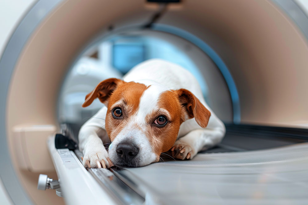
Radiographs reach their diagnostic ceiling the moment the clinical question outruns what superimposed, two-dimensional, low-contrast tissue imaging can answer. That happens more often than a busy general practice has time to catalog: a soft tissue mass that needs cross-sectional characterization, a suspected portosystemic shunt where flow matters more than form, a murmur whose severity lives in function rather than silhouette, a neurologic case where the lesion is anatomically invisible on plain film, or a surgical candidate whose approach depends on millimeter-level mapping. Recognizing the ceiling early, rather than after a second set of views comes back equivocal, is what separates a clean workup from a delayed diagnosis. The decision that follows, which modality and which specialist, is often the higher-value clinical call.
VESPECON exists to give referring veterinarians a concierge-level consultant on the other end of that call, without the friction of a formal transfer. Our board-certified radiologists provide diagnostic imaging interpretation, including radiograph and echocardiogram reads with structured follow-up recommendations, and our specialist network coordinates timely appointments when a case needs ultrasound, CT, MRI, or an on-site advanced workup. When the films are on your screen and the next step is unclear, reach out and we will consult on the case with you.
Key Takeaways
- Radiographs stay the right first-line tool for most small animal presentations, but a technically perfect study can still be functionally insufficient when the clinical question involves soft tissue contrast, three-dimensional anatomy, motion, or the central nervous system.
- Ultrasound is usually the next step rather than the last resort, adding real-time soft tissue architecture, Doppler flow, and image-guided sampling without ionizing radiation or, in most patients, general anesthesia.
- CT excels at bony detail, mass characterization, and vascular mapping; MRI is the gold standard whenever brain or spinal cord parenchyma is the question; echocardiography is the only way to assess cardiac function and chamber dimensions that a thoracic radiograph can only imply.
- The most useful outcome of advanced imaging is an interpreted study framed against a clear clinical question, and a pre-referral conversation often clarifies which modality fits before anyone books anesthesia.
Why Does a Normal Radiograph Not Rule Out Disease?
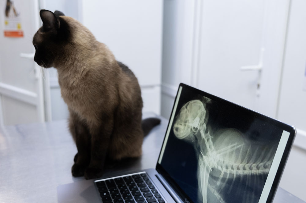
A normal radiograph does not rule out significant disease because plain film has real, predictable blind spots. Poor soft tissue contrast, two-dimensional superimposition, an inability to capture motion, and a brain and spinal cord that read as essentially opaque on film all define what radiographs can and cannot show, which is why an unremarkable study narrows the differential but never closes it.
The mechanics are worth keeping in front of mind. Two structures of similar density lying against each other blur into a single shadow. A swallowing disorder, a collapsing trachea, or vascular flow cannot register on a static image. And a patient with clear neurologic signs can have a completely normal-looking spine on plain film, because the cord itself is invisible.
None of this diminishes what radiography does well. Plain film is fast, widely available, and hard to beat for osseous detail, lung parenchyma, and the gross abdominal silhouette. It is the right tool for triage, for monitoring a known lesion over time, and for the majority of first-look questions a general practice fields in a day. The skill worth building is knowing where that tool stops, and communicating that ceiling is part of good diagnostic stewardship. Telling a colleague or client that films are “unremarkable so far” rather than “normal” keeps the door open to the next step when signs persist. Our tele-radiology exists in part for exactly that moment, when a study reads clean but the clinical picture says otherwise.
Where Does Ultrasound Fit Before You Reach for CT or MRI?
Ultrasound is the natural next step after radiographs for most soft tissue questions, and it is underused as a decision point rather than a last resort. It resolves the two problems plain film cannot solve on its own: it separates structures of similar density into distinct architecture, and it does so in real time, without ionizing radiation and usually without general anesthesia.
That combination makes it the highest-yield second study for a large share of general practice cases:
- Abdominal soft tissue architecture: liver, spleen, kidneys, adrenals, pancreas, and the layered wall of the GI tract, where a radiograph offers one undifferentiated silhouette.
- Fluid versus tissue: effusion, cystic versus solid masses, and the free abdominal fluid an AFAST scan can confirm in minutes during triage.
- Doppler flow: vascular patency, thrombi, and shunting, where the question is movement of blood rather than the shape of a vessel.
- Image-guided sampling: the decisive advantage, since a guided fine-needle aspirate or biopsy converts an image into a tissue diagnosis in the same visit.
- Serial monitoring: repeatable as often as the case demands, with no cumulative radiation and minimal patient cost.
The limits are equally specific, and they are what point you onward. Gas and bone reflect the beam, so aerated lung, the skull, and structures shielded by bone stay largely inaccessible. Deep or large-breed patients test penetration. Ultrasound does not map surgical margins or reconstruct a comminuted fracture the way CT does. And it is the most operator-dependent modality in the toolkit, which is precisely why the interpretation matters as much as the acquisition.
The practical rule: if the question is abdominal, soft tissue, or sampleable, ultrasound often answers it faster and more cheaply than CT. If the question is bony, vascular mapping for a surgical approach, thoracic parenchyma, or neurologic, ultrasound is the wrong stop and you should keep going.
When Is Echocardiography the Study You Need?
Echocardiography is the only modality that assesses cardiac function, and no thoracic radiograph substitutes for it. Radiographs answer a different question well: they show the cardiac silhouette, vertebral heart score, pulmonary vasculature, and the pulmonary edema that signals congestive failure. What they cannot show is why the heart looks that way, or how it is actually working.
Echo delivers what the silhouette only implies: chamber dimensions, wall thickness, valve morphology and regurgitant flow, systolic function, pressure gradients, and pericardial effusion. That distinction drives real decisions. Staging myxomatous mitral valve disease depends on chamber measurements that determine whether a patient has crossed into the stage where treatment changes. An asymptomatic murmur, a cat with suspected cardiomyopathy and a normal-looking silhouette, a congenital defect, or a pericardial effusion approaching tamponade all sit beyond what plain film can adjudicate.
The two studies are complementary rather than competing. Radiographs remain the better read on the lungs and on whether a patient is in failure; echo characterizes the heart itself. When a murmur, an arrhythmia, or an equivocal silhouette needs a cardiac answer, our tele-cardiology and tele-radiology services provide the interpretation with structured follow-up recommendations.
When Does CT Change the Diagnostic or Surgical Plan?
CT is the advanced modality reached for most often in small animal practice, and it changes the plan whenever three-dimensional detail or vascular mapping is the missing piece. Cross-sectional imaging removes overlapping shadows, soft tissue windowing sharpens contrast resolution, and scan speed keeps studies practical under anesthesia, which together explain when CT offers more than radiographs. CT angiography goes further, mapping vascular anatomy preoperatively with a precision plain film cannot approach, which matters enormously for a portosystemic shunt or a mass sitting on a major vessel.
Mass characterization and staging, head and neck disease where surgical margins matter, nasal and paranasal disease, complex fracture reconstruction, and vertebral lesion localization are the presentations where small animal CT imaging reliably changes the plan. A comminuted fracture that looks like a puzzle on two views becomes a solvable repair once it is reconstructed in three dimensions. A vertebral lesion gets pinpointed to the exact site before a decompressive approach is chosen.
CT has one important limit worth stating plainly: for the parenchyma of the brain and spinal cord itself, its contrast resolution trails MRI. It is the right call for bony lesions and vascular mapping, and the wrong first choice when intracranial or intramedullary soft tissue detail is the actual question.
When Is MRI the Right Choice Over CT?
MRI is the right choice whenever parenchymal detail of the brain or spinal cord is the clinical question. Superior soft tissue contrast without ionizing radiation, plus signal characterization across multiple sequences that separates inflammatory from neoplastic from vascular processes, is what makes small animal MRI the gold standard for that tissue. Just as importantly, it evaluates the spinal cord itself rather than only the bony canal around it, so a myelopathy gets assessed for cord signal change rather than inferred from vertebral shadows.
Seizure workups needing intracranial lesion evaluation, suspected brain tumor or encephalitis, myelopathy requiring cord signal assessment, peripheral nerve sheath tumors, and nasopharyngeal disease extending intracranially are the core indications for MRI for neurologic disease. Add hydrocephalus, brachial plexus evaluation, and middle ear disease with intracranial extension to that list.
The trade-offs are real: MRI asks for longer anesthesia, availability is more limited, and cost and positioning vary by body region. Neither modality is categorically superior, and the choice comes down to what you are trying to see.
| Consideration | CT | MRI |
| Best for | Bone, vascular mapping, mass staging | Brain and spinal cord parenchyma |
| Soft tissue contrast | Better than plain film | Superior, across multiple sequences |
| Ionizing radiation | Yes | No |
| Scan and anesthesia time | Faster, shorter anesthesia | Longer anesthesia required |
| Availability | Wider | More limited |
When a case points toward the nervous system, our board-certified radiology and neurology specialists can talk through whether MRI is the study that will actually answer the question.
What If the Clinical Question Is About Motion?
When the pathology is the movement itself, no still image will answer the question. Fluoroscopy provides real-time visualization of a dynamic process, from dysphagia and aspiration workup to dynamic tracheal collapse, esophageal motility, and guidance for interventional procedures. A swallowing study can pinpoint which phase of the swallow is affected, catch aspiration in the act, and separate pharyngeal from esophageal dysfunction. A collapsing trachea shows its true behavior only under the physiological loading of an actual breath.
Fluoroscopy complements cross-sectional imaging rather than competing with it. A dog with esophageal disease may need fluoroscopy to characterize function and CT or endoscopy to characterize structure. Function and form are two different questions, and the smart workup asks them in the right order.
When Do You Need to See the Lumen?
You need a contrast study when the question is about the lumen itself: mucosal detail, luminal patency, or the structural relationships of the gastrointestinal tract, particularly when endoscopy is not immediately available. Suspected partial obstruction, esophageal stricture, gastric emptying questions, and ruling perforation in or out are the indications for a positive contrast gastrointestinal study, whose diagnostic value rises and falls with patient preparation, contrast agent selection, and projection timing. When perforation is on the table, water-soluble contrast is the safer agent choice. When a partial obstruction refuses to declare itself on plain film, the passage of contrast over timed projections often tells the story.
The interpretation is only as good as the technique feeding it, which is one more reason a shared read helps. When a contrast study raises as many questions as it answers, a specialty consultation on a difficult case can help sort the ambiguous findings from the actionable ones.
How Does PET-CT Add to Standard Imaging?
PET-CT adds metabolic information to anatomic detail. Layering glucose metabolism onto cross-sectional imaging lets PET-CT flag active tumor tissue, treatment response, and occult metastatic spread that falls below the size threshold CT can resolve on its own. In oncology, that extra layer can change the staging conversation entirely.
The indications where it earns its cost tend to be specific: staging a known malignancy where metastatic burden will steer the plan, post-treatment surveillance where anatomic imaging cannot tell scar from residual viable tumor, and equivocal CT findings where metabolic activity would tip the decision. The limits deserve equal honesty. Availability is restricted to a small number of academic and specialty centers, cost runs well above CT or MRI, and the evidence base for specific veterinary tumor types is still maturing.
PET-CT is not the only functional study worth knowing. Nuclear scintigraphy remains useful in a narrow set of questions, including portosystemic shunt confirmation, thyroid imaging, and bone scans that localize an occult lameness, though it is similarly limited to referral centers with the licensing to handle radioisotopes. For cases where a metabolic or functional answer would change management, our oncology and radiation oncology advisors can help weigh whether the added information is worth the trip.
Frequently Asked Questions About Advanced Imaging
When should I go straight to CT instead of starting with ultrasound?
Go straight to CT when the question is bony, vascular, thoracic, or surgical. Ultrasound cannot penetrate aerated lung or bone, will not map margins for a surgical approach, and will not reconstruct a fracture. If the question is abdominal soft tissue, an effusion, or a mass you want to sample, ultrasound usually answers it faster, cheaper, and without general anesthesia, and it may make CT unnecessary altogether.
How do I decide between CT and MRI for a given case?
It comes down to the tissue in question. If you need bony detail, vascular mapping, or mass staging, and your patient tolerates only short anesthesia, CT usually wins. If the parenchyma of the brain or cord is the question, MRI is the study that will actually show it. When it is genuinely unclear, a quick pre-referral conversation settles it faster than a repeat set of films.
How should I explain a normal radiograph to a client when I still suspect disease?
Frame the study as “unremarkable so far” rather than “normal,” and explain plainly that plain film has known blind spots. It sees bone well but resolves soft tissue poorly, flattens three dimensions into two, cannot capture motion, and does not show the brain or spinal cord. That honest framing keeps the client prepared for a next step if signs persist, and it protects the relationship when advanced imaging later finds what radiographs could never have shown.
What information makes an imaging referral most useful?
A clear clinical question. Tell the radiologist what is already known, what the differential looks like, and what the imaging specifically needs to answer. A note that says “left-sided head tilt, suspect middle ear versus intracranial extension, need to know if the process crosses the tympanic bulla” produces a far more actionable report than “evaluate head.” The interpretation can only be as sharp as the context you provide with the study.
Let’s Talk Through the Case Before You Refer
The most useful outcome of advanced imaging is never just the image. It is the interpreted study, read in the context of your clinical question, with a clear recommendation for the patient’s next step. That is the part a specialist adds, and it starts well before anyone books a scan.
Anesthesia duration, positioning that varies by body region, and cost are the practical realities worth settling before the study rather than after. We are here to help you decide whether imaging is indicated, which modality fits the question, and how to prepare the patient and the client. If you have a case that warrants the conversation, collaborate with us on new and repeat cases, and we will think it through alongside you. You can also start a pre-referral case discussion whenever a study leaves the next step unclear.



Leave A Comment