
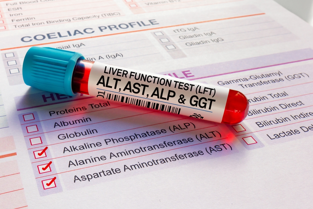
An elevated liver enzyme is a finding, not a diagnosis, and the pattern usually tells you more than the magnitude. ALT and AST reflect hepatocellular leakage, ALP and GGT track cholestasis, and their differing half-lives are what make recheck timing a clinical decision rather than a default interval. A mild ALT increase in a well dog may reasonably earn a four-week recheck, while any ALP elevation in a cat warrants attention now, because feline patients have no steroid-induced isoenzyme. Many elevations also trace back outside the liver, to endocrine disease, drug induction, or a reactive hepatopathy driven by GI or pancreatic inflammation. Making that call early is the difference between a repeat panel in a month and a bile acid and imaging workup this week.
VESPECON gives your practice on-demand access to board-certified internists who will work a challenging enzyme pattern through with you and help develop a plan for imaging, medications, or surgery. Abdominal ultrasound is usually the first look at the liver itself, and when a case moves on to radiographs or CT, our radiologists read those studies, with STAT options available. Get in touch with us when a set of liver values is not adding up and you want a specialist’s read before the next recheck.
Liver Disease in Dogs: The Highlights
- ALT and AST mark hepatocellular leakage while ALP and GGT mark cholestasis, and the relationship between the two groups narrows the differential faster than the multiple of any single value.
- Most ALP elevations in dogs trace back to drug induction, endocrinopathy, or reactive change rather than primary hepatobiliary disease.
- Urgency comes from function, not enzyme activity: albumin, glucose, BUN, cholesterol, bilirubin, and bile acids tell you whether the organ is failing.
- Imaging can’t separate nodular hyperplasia from neoplasia, so cytology or histopathology remains the endpoint for focal lesions and for diffuse parenchymal change that won’t resolve.
What Do the Four Liver Enzymes on a Chemistry Panel Measure?
The four enzymes split into two functional groups. ALT and AST sit inside hepatocytes and leak out when those cells are injured, so they mark damage to the parenchyma. ALP and GGT are anchored to biliary membranes and rise when bile flow is impaired. Which of the two groups dominates sets the direction of the workup.
ALT and AST: Hepatocellular Leakage
ALT is the most liver-specific of the four in dogs, with a serum half-life on the order of two to three days, which makes it workable for tracking recovery after an acute insult. AST comes from hepatocyte mitochondria and cytosol but also from muscle and erythrocytes, so a rise without a matching ALT should send you to a CK and a look at the sample for hemolysis before the liver enters the conversation.
Magnitude is a poor proxy for severity. A dog who found something hepatotoxic in the garage can run four figures and normalize inside a month, while a Doberman holding a three-fold elevation with a slipping albumin is in considerably more trouble. You’re reading a trend line, not a snapshot, and two or three serial panels on the same analyzer say more than one dramatic number. When a borderline pattern refuses to settle and you’re weighing whether to sample, a clinical pathology consultation costs less than a biopsy the case didn’t need.
ALP and GGT: Cholestasis and the Specificity Problem
ALP and GGT live on the membranes of bile duct cells, so they climb when bile flow slows down, and that pattern steers the workup toward liver and gallbladder disorders instead of toward damaged liver cells. In dogs, ALP is the enzyme most often flagged and the least informative on its own. Three isoenzymes feed it: a hepatic form, a bone form that runs high in growing patients and in bone-active disease, and a corticosteroid-induced form that answers to endogenous cortisol as readily as to prednisone. Phenobarbital induces it too, and several breeds, Scottish Terriers among them, carry benign elevations that never declare anything.
GGT is the more discriminating partner. When it climbs alongside ALP, the odds shift toward biliary disease, a mucocele, or a mass distorting the biliary tree rather than simple induction. Isoenzyme fractionation gets discussed more often than it gets ordered, and the interpretation rather than the assay is where the value usually sits, which is one reason it keeps surfacing in the continuing education for veterinary teams we build.
| Enzyme | Main source | Approximate canine half-life | Climbs with | Chief confounder |
| ALT | Hepatocyte cytosol | 2 to 3 days | Hepatocellular injury | Modest contribution from severe myopathy |
| AST | Hepatocyte mitochondria, muscle, RBC | Hours | Hepatocellular injury, myopathy, hemolysis | Non-hepatic sources; pair it with CK |
| ALP | Biliary membrane, bone, steroid-induced isoenzyme | About 3 days | Cholestasis, drug induction, bone activity | Least specific value on the canine panel |
| GGT | Biliary epithelium | Days | Cholestasis, biliary hyperplasia | Colostrum ingestion in neonates |
What Causes Liver Enzyme Elevations in Dogs?
Two categories cover nearly everything. Primary hepatobiliary disease means the injury starts in the liver: inflammation, copper accumulation, toxin exposure, a vascular malformation, or neoplasia. Secondary change means the liver is reacting to trouble somewhere else, and in general practice that second group accounts for the majority of abnormal panels.
Primary Hepatobiliary Disease
- Inflammatory and infectious: Inflammation of the liver tissue itself, canine hepatitis, can trace back to an infectious cause the core vaccine series already covers, to copper accumulating in the liver of a predisposed breed, or to a drug or toxin the liver was handed to process. Leptospirosis belongs on the same list and deserves testing early in an acute icteric case.
- Vascular: In a young dog with stunted growth, wobbliness or disorientation an hour after eating, and values that never quite settle, portosystemic shunts send blood around the liver rather than through it, and the correction is anatomic rather than medical.
- Toxic and drug-associated: Xylitol, aflatoxin, sago palm, blue-green algae, and acetaminophen produce acute hepatocellular necrosis. Idiosyncratic reactions to carprofen, azathioprine, lomustine, and trimethoprim-sulfa show up without warning and resolve on withdrawal if you catch them.
- Copper storage: Bedlington Terriers, Labradors, Dobermans, Dalmatians, and West Highland White Terriers accumulate hepatic copper, and quantitative copper on a biopsy sample is the only way to confirm it and dose chelation sensibly.
- Neoplastic and structural: Hepatocellular carcinoma, biliary carcinoma, metastatic disease, and gallbladder mucocele all present as enzyme changes long before they present as clinical illness.
Survey radiographs still earn their place early, since hepatic size, silhouette, and mineralization shape what you look for on ultrasound. When a study is equivocal, radiology study interpretation is worth pursuing before a family commits to anesthesia and a sampling procedure.
Secondary and Reactive Hepatopathy
Cushing’s disease, pancreatitis, inflammatory bowel disease, and long courses of steroids or anticonvulsants all push enzymes upward without the liver being the sick organ, which is why liver disease in dogs gets diagnosed from the whole patient and not from one line on a panel. Vacuolar hepatopathy is the histologic signature of that group, and it’s reversible when the driver is addressed.
The portal drainage explains much of it. Pancreatic and intestinal inflammation delivers cytokines and bacterial products straight to the sinusoids, and the hepatocytes respond. Sepsis produces functional cholestasis with hyperbilirubinemia and no obstruction at all. Right-sided congestive failure backs pressure into the hepatic veins and gives you a big liver with modest enzyme changes, and when the panel and the radiographs point that direction, an echocardiogram and an ECG answer the question faster than more liver testing does. Both of those studies come back to us for interpretation.
Phenobarbital deserves its own note. Induction is expected and by itself means very little. What matters is a disproportionate ALT climb, a falling albumin or BUN, rising bile acids, or a patient who has stopped feeling well.
When Does an Elevation Warrant a Workup Now Instead of a Recheck?
Urgency comes from function and clinical status rather than from the multiple of the reference interval. A well dog with an isolated two-fold ALT can reasonably wait four weeks. Hypoalbuminemia, hyperbilirubinemia, hypoglycemia, prolonged clotting times, icterus, weight loss, or altered mentation move the same patient into a same-week workup.
| Pattern | Likely interpretation | Reasonable next step |
| Mild isolated ALT, well dog, no medications | Transient or reactive injury | Fasted recheck in about four weeks |
| Isolated ALP in an older dog with PU/PD | Induction or endocrinopathy | Screen the adrenal axis before sampling the liver |
| ALT and ALP both rising across serial panels | Progressive hepatopathy | Bile acids, abdominal ultrasound, plan for sampling |
| Any hyperbilirubinemia or visible icterus | Cholestasis, hemolysis, or obstruction | PCV and slide review, then imaging this week |
| Low albumin, BUN, cholesterol, and glucose | Loss of functional mass | Urgent; coagulation panel before anything invasive |
| Marked acute ALT with toxin exposure | Acute hepatocellular necrosis | Treat now, characterize later |
Three reflexes are worth resisting. Empiric glucocorticoids in an undiagnosed hepatopathy blur the histology and can worsen an infectious or copper-associated case. Never sample the liver before assessing coagulation, since cholestatic patients lose vitamin K dependent factors and platelet function can be poor even when the count looks reassuring. And don’t hand icterus to the liver until a PCV and a blood smear have ruled out hemolysis, because pre-hepatic and post-hepatic causes sit on either side of that finding. When a pattern is moving and the next decision is a bile acid pair versus a sampling procedure, reach out and we’ll take the case apart with you first.
How Do Feline Liver Patterns Differ From What You See in Dogs?
Cats make no steroid-induced ALP, so any elevation carries weight, and liver disease in cats can travel from a few skipped meals to hepatic lipidosis inside of a week. Feline ALP also has a shorter half-life and a lower hepatic concentration, which means a small number means more.
GGT is the discriminator worth watching in a mixed caseload. It rises with cholangitis and biliary disease but stays flat or nearly flat in lipidosis, so a marked ALP with an unremarkable GGT in an anorectic cat points hard toward lipid accumulation. Triaditis, the cholangitis, pancreatitis, and enteropathy cluster, keeps the workup from stopping at the liver. In a cat, anorexia is the finding that sets the clock, not the enzyme.
What Does the Workup Look Like After the First Abnormal Panel?
The first move is a history and a full-panel review of liver function tests rather than an image. Medication and supplement lists, toxin access, breed, body condition, and the rest of the chemistry, including albumin, bilirubin, glucose, BUN, and cholesterol, decide whether this patient needs a repeat panel next month or a function test this week.
Initial Evaluation and Monitoring
Pre- and postprandial bile acids answer the functional question that enzymes can’t, and fasting ammonia adds to it when encephalopathy is on the table. A urinalysis with specific gravity, a UPC if protein shows up, and adrenal testing when the clinical picture supports it round out the first pass. Mild, isolated hepatocellular elevations in a well dog reasonably run on a four-week interval; a stable drug-induced ALP in a dog who feels fine can stretch to eight or twelve weeks. Keep the samples on the same analyzer so the trend means something.
Advanced Diagnostics for Persistent or Severe Elevations
A nodule spotted on an older dog’s abdominal ultrasound is not automatically liver cancer, because benign nodular hyperplasia looks much the same on the screen and only sampled cells answer the question. Ultrasound sorts diffuse from focal disease, characterizes the gallbladder wall, and screens the portal vasculature. CT angiography maps anomalous vessels and stages a hepatic mass with far more confidence than ultrasound offers.
Cytology and histopathology disagree more often than most of us expect in diffuse hepatic disease. Aspirates are reliable for lipidosis, lymphoma, and obvious vacuolar change, and unreliable for chronic hepatitis, fibrosis staging, and copper quantification, all of which need tissue. Coagulation assessment comes first regardless of technique. Wedge and laparoscopic samples give better architecture than needle cores, and we coordinate transfer to a specialty hospital when a case needs laparoscopic or surgical biopsy, then stay on the case for the histopathology conversation afterward.
How Does Treatment Follow the Diagnosis?
Treatment is cause-directed, and outcomes track how well the primary problem gets controlled. Withdraw the offending drug, chelate copper when quantification supports it, treat the infection, manage the endocrinopathy, correct the shunt surgically. Nonspecific hepatoprotectants support that plan; they aren’t a substitute for one.
Cause-Directed Therapy and Supportive Care
With acute liver disease, most of the treatment is scaffolding while the organ rebuilds itself: intravenous fluids, glucose and electrolyte correction, antioxidant support, and control of the ammonia buildup that clouds a dog’s mentation. Doxycycline covers leptospirosis while titers are pending. Bacterial cholangitis wants culture-guided therapy, ideally from bile. N-acetylcysteine has a role in acetaminophen toxicosis and other acute hepatotoxic events, and SAMe with silybin carries the antioxidant load afterward.
Beyond that, the choices narrow to the diagnosis in hand. Ursodiol improves bile flow in non-obstructive cholestasis. Immunosuppression belongs to biopsy-confirmed idiopathic chronic hepatitis and not to a suspicion. D-penicillamine plus dietary copper restriction manages storage disease. Lactulose, dietary protein modification, and gut-directed antimicrobials handle encephalopathy, and parenteral vitamin K is worth giving before any procedure in a chronically cholestatic patient.
When Nutritional Support Becomes the Intervention
Calories are therapy in hepatic disease, and protein restriction is overused. Unless a patient is encephalopathic, keep high-quality protein in the bowl and meet resting energy requirement, because negative energy balance drives catabolism and worsens the very problem you’re treating. When a pet has refused food for days and calories cannot wait for appetite to return, an esophagostomy tube goes in under brief anesthesia and carries full nutrition and medications at home for weeks, without a wrestling match at every meal. Appetite stimulants such as mirtazapine or capromorelin bridge a short gap; they don’t replace a feeding plan in a patient who has been anorectic for the better part of a week.
What Referring Veterinarians Ask Us About Liver Panels
Do normal bile acids rule out significant hepatic disease?
They don’t. Bile acids assess functional mass and portal circulation, and the canine liver holds enough reserve that early chronic hepatitis or a low-grade copper accumulation can sit behind a normal result. They’re also redundant in a patient who is already icteric, since hyperbilirubinemia has answered the same question. Use them where they discriminate: a persistent enzyme elevation without icterus, a small-breed dog with suspicious neurologic signs, or a decision point about whether to move toward tissue.
How long can a phenobarbital-treated dog’s ALP be allowed to climb?
Induction alone isn’t a reason to change course, and many well-controlled epileptics run an elevated ALP for years. The findings that should prompt action are a disproportionate ALT rise, a falling albumin or BUN, an increase in bile acids, or a dog who has become lethargic, ataxic beyond baseline, or inappetent. Check a serum phenobarbital concentration alongside the panel, and consider transitioning to an anticonvulsant with a different metabolic profile if the trend continues.
Do liver enzymes need to normalize before anesthesia?
Enzyme activity by itself isn’t a contraindication. What determines anesthetic risk is functional capacity: albumin, glucose, coagulation times, platelet count, and the patient’s ability to metabolize what you give them. A dog with a mild isolated ALT and normal function usually tolerates a routine protocol. A hypoalbuminemic, coagulopathic, or hypoglycemic patient needs the plan rebuilt around those deficits, with dose reductions, glucose support, and blood products available.
When to Bring a Specialist Into a Liver Workup
Most liver cases don’t declare themselves on the first panel. The hard part is deciding which patient repeats a chemistry in four weeks and which one needs bile acids, imaging, and a sampling plan this week, and that call is easier to make out loud with someone who sees these patterns every day. VESPECON keeps board-certified internists, radiologists, and cardiologists reachable while a case is still in front of you, from the first odd value through sampling, transfer, and long-term monitoring. When the numbers stop adding up, start a consultation request and we’ll take the pattern apart together.



Leave A Comment